


Electron Probe MicroAnalysis (EPMA) is a non-destructive technique to determine chemical composition of small amounts of solid materials. A focused beam of high-energy electrons hits the sample and generates characteristic x-rays corresponding to the elements present in the material. The beam current is typically between 10-100nA; much more intense than SEM. This produces a higher count rate, thus improving precision, accuracy and detection limits (~100s ppm).
Characteristic x-rays are produced by inelastic collisions of the incident electrons with electrons in the inner shell of atoms in the sample. When an inner electron is ejected from its orbit, it leaves a vacancy which is filled by a higher-shell electron dropping into the vacancy. The higher-shell electron sheds energy in the form of an x-ray, which is characteristic of the particular element.
The electron microprobe uses Wavelength Dispersive Spectroscopy for precise quantitative chemical analysis. A small fraction of x-rays escape from the sample, reach a crystal of known lattice spacing, and is diffracted into the crystal at a particular angle (the Bragg angle). Thus, a WDS spectrometer is tuned into one particular wavelength and sits for a specified period of time and counts the number of x-rays coming through the crystal at that angle.
EPMA is a standard-based technique. For precise analysis, similar standards of known composition are used. Using the same settings on both standards and unknown samples, you can determine the weight fraction of the unknown. A matrix correction (e.g. ZAF or PAP) is then applied to determine the composition of the unknown.
Chemical analysis by EPMA also provides textural context. Small-scale variation within a sample can be determined accurately. The activation volume excited by the beam is typically on the order of 2µm, even though the beam itself is less than 1µm.
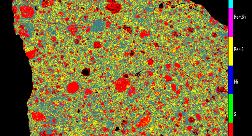
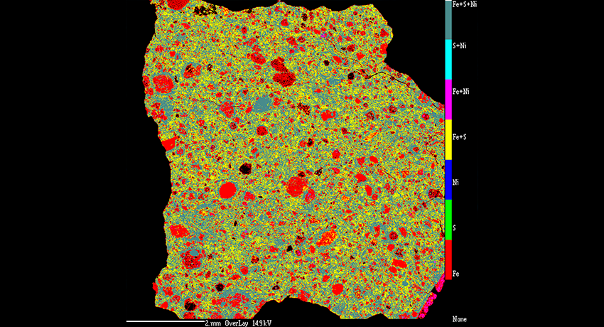
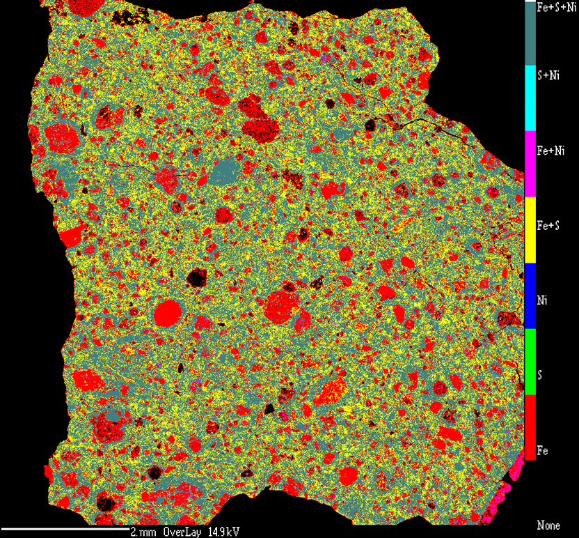
A compositional map of a chondritic meteorite.
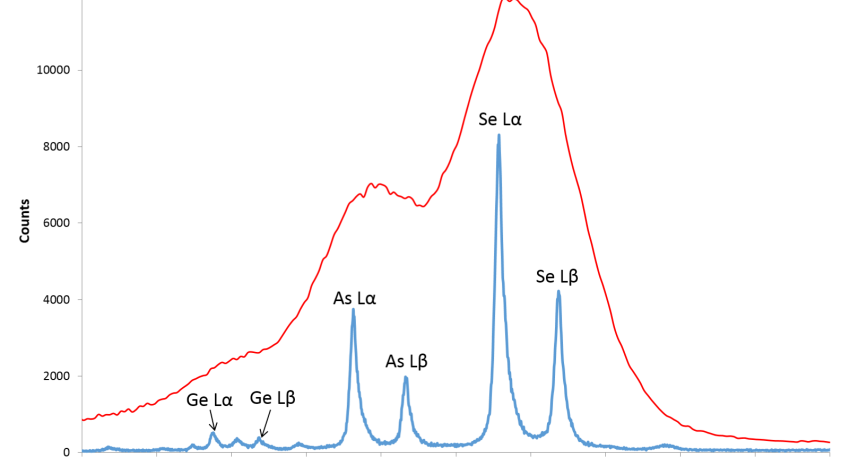
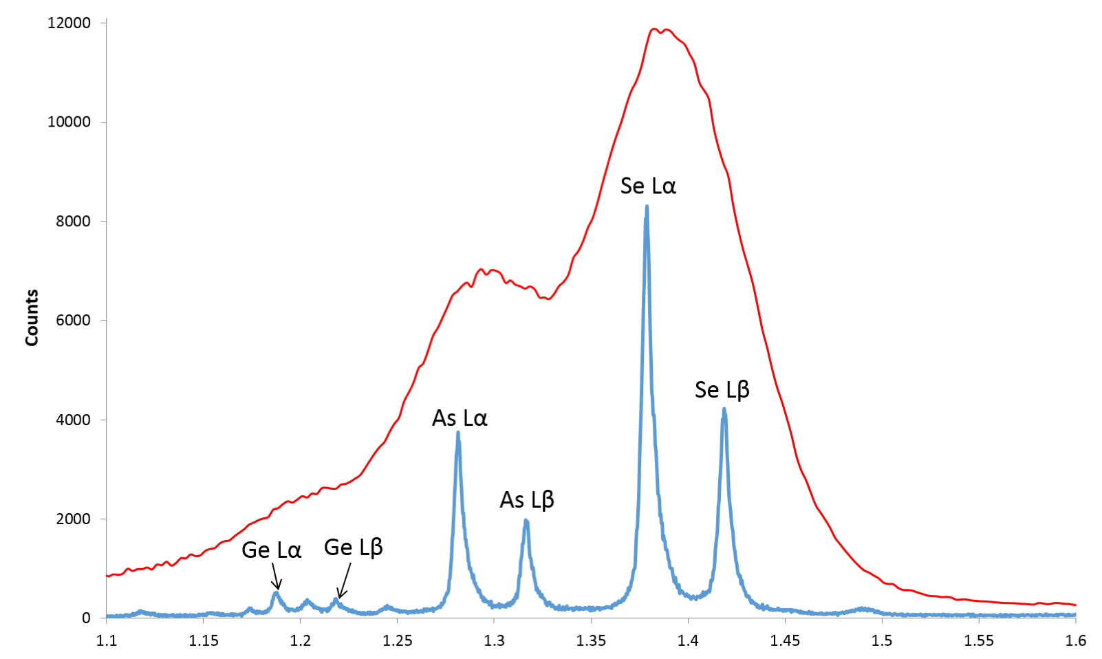
The same conditions were used for both spectra. EDS has significant peak overlaps between Selenium (Se), Arsenic (As) and Germanium (Ge). However, WDS can clearly distinguish between each of the elements.

Read all the details about EPMA characterization technique at Penn State. Areas include:
- CAMECA SX-Five
- LaB6 filament
- Integrated Bruker EDS software
- Five WDS spectrometers
- Elemental coverage Be and up
- Crystals available: LIF, PET, TAP, PC
- SE2 and BSE detectors
- Large catalog of standards
- Accommodates samples of various sizes, limited to 18mm tall
Cameca SX Five Capabilities
- Simultaneous X-ray (WDS and EDS), SEM and BSE imaging
- High sensitivity (few tens of ppm) from sub-micron volumes
- Elemental detection from Beryllium (Be) to Uranium (U)
- Optimized vacuum system for low-level analysis of carbon and boron
- Three high sensitivity/high resolution WD spectrometers; Two 4-crystal high Resolution Spectrometers
- Multilayer/thin film analysis
- Monazite age dating
- LaB₆ Electron Source for maximum versatility
For more information about this instrument, visit their website: CAMECA Science & Metrology Solutions