

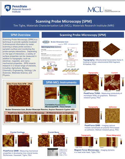
An Atomic Force Microscope (AFM) provides 3-dimensional topographic information about a sample by probing its surface structure with a very sharp tip. The tip is scanned laterally across the surface, and the vertical movements of the tip are recorded and used to construct a quantitative 3-dimensional topographic map. The lateral resolution of the image can be as small as the tip radius (typically 1-15 nm), and the vertical resolution can be on the order of angstroms.
Technique Advantages
- Quantitative topographical information at high lateral resolution
- Little or no sample prep in many cases
- Little to no harm to sample
- Applicable to conductive and insulating materials
Typical Applications
- High-resolution surface profilometry
- Surface roughness measurements
- Microstructural studies of metallic, ceramic, semiconducting and polymeric materials
- Defect and failure analysis
- Semiconductor device structural analyses
- Surface cleaning and polishing studies
- Phase separation in polymers
- Investigation of local mechanical properties (i.e. stiffness, adhesion, friction)
- High-resolution imaging of biological samples
- Studies of nano-scale forces
- Contact Mode, Lateral Force Microscopy.
- Piezo Response Microscopy.
- Conductive AFM, Scanning Capacitance Microscopy.
- Tapping Mode w/ Phase Imaging.
- Electrostatic Force Microscopy, Magnetic Force Microscopy,Kelvin Probe Force Microscopy.
- PeakForce Tapping w/ ScanAsyst.
- PeakForce Quantitative Nanomechanical Mapping.
- PeakForce TUNA • PeakForce Kelvin Probe Force Microscopy • Dimension heater/cooler stage enables AFM at two different temperature ranges:
- -35ºC up to 100ºC
- Room temperature up to 250ºC
- High resolution imaging of biological samples in buffer solution (tissues, cells, viral particles, hydrogels applications, biomedical devices, and individual biomolecules).
- High resolution investigation of local mechanical properties (i.e. stiffness, adhesion, friction) of biological samples.
- Time resolved imaging of biological samples up to 98 scan lines per second.
- Studies of nano-scale forces between ligands in the single molecule range (Molecular interaction mapping, Single Molecule Force Spectroscopy (SMFS).
- Correlated fluorescence and Atomic Force Microscopy (AFM) operation for high-resolution data collection and functional targeting of samples of interest (e.g. tissues, surfaces, cells).
- High resolution video-rate imaging at up to 625 lines per second which leads to at least 10× faster imaging than current “fast scanning” AFMs.
- Captures data directly to a video format, not individual image frames.
- Capture images at high pixel density (512×512) in just over a second, or reduce scan lines to achieve video rates >10 fps.
- blueDrive photothermal excitation makes video-rate AFM simple and stable.
- Scanner is extremely robust and fully sealed against liquid spills for complete reliability.
- Motorized laser and detector alignment make quick work of instrument set-up.